Many times, we do not know exactly the differences between computed tomography (CT) and magnetic resonance imaging (MRI). If you are one of them then you are in right place, below we will discuss MRI vs Xray. Their utilization, benefits and in what circumstances these scan tests should be used.
CT uses ionizing radiation; that is, X-rays, to obtain detailed images of internal organs, bones, soft tissues, and blood vessels, while MRI uses a strong magnetic field, radio waves, and a computer to produce detailed images of internal structures of the body.
Thus, CT images are cross-sections that can be reconstructed in multiple planes and even processed in three dimensions that can be viewed on a computer monitor, printed on a plate or paper, or transferred electronically. It is a fast, painless, precise and non-invasive technique. In emergencies, it can identify injuries, strokes and internal bleeding fast enough to help save lives.
In fact, doing a CT study is faster, since it takes a few seconds, it is more comfortable and less noisy than magnetic resonance imaging, since the cavity (gantry) into which the patient enters is wider and shorter, so it does not generate a feeling of claustrophobia.
For its part, MRI images are clearer, more detailed and on certain occasions, can identify and characterize diseases with greater probability than other diagnostic imaging methods. MRI is a non-invasive procedure and, unlike CT, does not use ionizing radiation.
The examinations are longer, since depending on the test they usually last between 10 and 45 minutes, and the cavity (gantry) where the patient is inserted is narrower and longer, so it can give a feeling of being overwhelmed. “It also makes a loud and strange noise, like hitting, which makes you think that the machine has broken, but it is normal and should not worry you”, details our specialist.
CT scanning can study many organs at once and is generally the best method for handling many clinical situations and detecting various types of cancers, as the images allow the radiologist to confirm the presence and determine the size and location of cancers. a tumor, and see the pathology that affects different organs and musculoskeletal structures. It is also sometimes used to complete MRI or ultrasound studies.
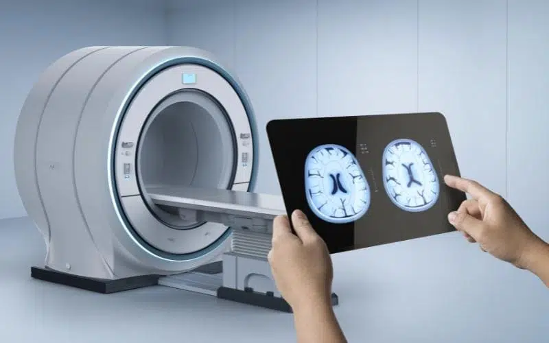
For its part, MRI is used, above all, to evaluate a specific organ in a more targeted way, such as the brain and spinal cord, some joint, spine, liver, heart and pelvis – rectum, gynecological, and prostate. It is also used to complement the study of some parts of the body that have been evaluated by other techniques (such as CT or ultrasound). Unlike CT, it can be used to study an unborn child inside the womb.
According to our radiologist, practically all the organs of the body can be studied with both techniques, although depending on the doctor’s clinical suspicion, the patient’s situation and in agreement with the radiologist, one or the other will be more indicated. For example, the brain is seen well with both techniques, but if you have suffered a blow to the head, it is preferable to study the patient with a CT; if the suspicion is something else, for example, a tumor, an MRI might be preferable.